Introduction
Introduction
What colours are seen by people with abnormal colour vision?
Some people have difficulty perceiving colours correctly. Colour is used widely today as a means of encoding information. In some occupations, such as flying aircraft, the recognition of colours on electrical devices is important. For this reason, much is known about how accurately people recognise colours on such devices. However, little is known about the perception of surface colours, like those displayed on computer screens.
Professor Barry Cole and his colleagues, Ka-Yee Lian and Carol Lakkis, were interested in learning more about this so that the design of colour-coded displays could be improved. They collaborated with statistician Dr Ken Sharpe on this project.
The key question was: How accurately can people with abnormal colour vision name surface colours?
Background
Clinical vision tests
Four clinical vision tests were used. These were the Ishihara test, the Farnsworth D15 test, the Richmond HRR test and a test using the Type I Nagel anomaloscope. The results of the four clinical colour vision tests were used together to identify the type of colour vision deficiency each participant had. All participants were classified as having one of six colour vision deficiencies.
Reduced or impaired sensitivity to the green end of the spectrum:
- Deuteranomaly (Mild)
- Deuteranomaly (Moderate)
- Deuteranopia (Severe)
Reduced or impaired sensitivity to the red end of the spectrum:
- Protanomaly (Mild)
- Protanomaly (Moderate)
- Protanopia (Severe)
See the Glossary for more details of these impairments.
-
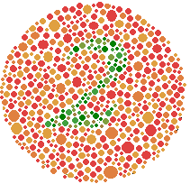
The Ishihara test for colour vision deficiency is relatively well-known. It was first published by a Japanese academic, Dr. Shinobu Ishihara, in 1917. It is made up of a series of images of circles. Each circle contains a large number of different sized and different coloured dots. Some dots are coloured in such a way to distinguish a number. Colour blind people may not be able to see the number or may see a number different from that seen by people with normal vision. The Ishihara test does not have images to detect "blue-yellow" colour vision deficiencies.
-
The Farnsworth D15 test uses 16 coloured disks that make up a complete colour circle. A tester places the first reference disk down and then asks the person being tested to place down the next closest colour. They are then asked to put down the next closest colour, and so on, until all the disks are placed in a line. A person with colour vision deficiency will place the disks in an order that is different from the order used by people with no deficiency. The particular order of the disks can be used to diagnose the type of colour vision deficiency. The Farnsworth D15 can also be used to classify the degree of colour vision deficiency.

-
The Richmond Hardy-Rand-Rittler (HRR) test, like the Ishihara test, is made up of a series of images created from patterns of dots. The Richmond HRR test has simple symbols, rather than numbers, encoded in colour within the image.
When the test is used with adults, they are asked to trace the symbol in the image with a camel's hair brush. There are four practice images, and 20 test images.
Adults who get the first 6 test images correct have normal colour vision; the pattern of errors (if any) on the set of test images can be used to diagnose the nature of the colour vision deficiency.
-
The Nagel anomaloscope is an optical instrument that a person looks into to see a coloured circle. The top and bottom halves of the circle are coloured differently, and the observer's task is to adjust two knobs to make the bottom half of the circle match the top half in colour and brightness.
The two controls on the anomaloscope allow the observer to adjust the amount of yellow light, and the ratio of red to green. The type of colour vision deficiency (if any) can be diagnosed from the way adjustments are made.
Colour naming task
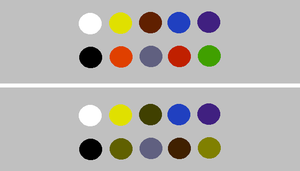
The colour naming task was developed especially for this study. It was devised to test people's ability to name ten different surface colours correctly. The colours were red, orange, brown, yellow, green, blue, purple, white, grey and black. A group of experimenters with normal colour vision chose the colours so that they were an unambiguous instance of the colour they wished to test.
The colour naming task used 6 A4 cards with the ten colours. The colours were displayed in different ways on each card; three cards used dots with diameters of 17mm, 7mm and 1.9mm. The other three cards used 20mm lines which were 3.5mm, 1.9mm and 1mm wide. The number of errors made out of 10 was recorded for each card. Participants judged each card twice.
Publication
In 2006, a paper describing the study's findings was published:
Cole, B. L., Lian, K. Y., Sharpe, K., & Lakkis, C. (2006). Categorical color naming of surface color codes by people with abnormal color vision. Optometry and Vision Science, 83(12), 879-886.
The publication includes a group of 20 participants with normal colour vision but these were not the main focus of analysis; these are not considered in the data provided here.