Analysis
Summary
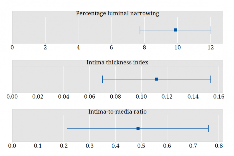
Brian, Lek and their colleagues found that there were more abnormalities in radial arteries (RA) than in internal thoracic arteries (ITA). They also found differences between the arteries for a number of characteristics measured in morphometric analysis. They cautioned that care should be taken in using the radial artery for CABG surgery in some clinical subpopulations.
These figures show estimates and 95% confidence intervals for the mean difference (RA – ITA) for three indices of severity of arterial disease:
Questions to consider
- Produce a summary table to describe the characteristics of the study participants.
- Produce visual displays to describe the results of the morphometric analysis of ITAs and RAs.
- Describe the distribution of each of the three main indices of severity of disease (%LN, ITI, IMR) using appropriate visual displays.
- Compare ITAs and RAs on the three main indices of severity of disease. Summarise your results with a confidence interval.
- Compare ITAs and RAs on the width and area measures obtained from the morphometric analyses. Calculate and plot confidence intervals to summarise your results.
- Which risk factors predict each of the three main indices of severity of disease? Conduct separate analyses for ITAs and RAs.
- Which risk factors predict the arterial abormalities identified by the pathologist? Examine one of the abnormalities and conduct separate analyses for ITAs and RAs.
Definition of variables in data file
| Age | Patient age in years |
|---|---|
| Gender | Sex: 1 = Male, 0 = Female |
| Diabetes | Presence of diabetes mellitus: 0 = no, 1 = yes |
| Ever smoked | History of cigarette smoking: 0 = never, 1 = ever |
| PVD | Presence of Peripheral vascular disease (PVD): 0 = no, 1 = yes |
| CVD | Presence of Cerebrovascular disease (CVD): 0 = no, 1 = yes |
| Hypercholesterolemia | Presence of Hypercholesterolemia: 0 = no, 1 = yes |
| RA intimal abnormality | Radial artery – Intimal abnormalities: 0 = normal, 1 = intimal thickening, 2 = atherosclerosis |
| RA medial calcification | Radial artery – Medial calcification: 0 = no, 1 = yes |
| ITA intimal abnormality | Internal thoracic artery – Intimal abnormalities: 0 = normal, 1 = intimal thickening, 2 = atherosclerosis |
| RA % luminal narrowing | Radial artery – Percentage of luminal narrowing |
| RA Intimal thickness index | Radial artery – Intimal thickness index |
| RA Intima-to-media ratio | Radial artery – Intima-to-media ratio |
| ITA % luminal narrowing | Internal thoracic artery – Percentage of luminal narrowing |
| ITA Intimal thickness index | Internal thoracic artery – Intimal thickness index |
| ITA Intima-to-media ratio | Internal thoracic artery – Intima-to-media ratio |
| RA DLI | Radial artery – Diameter of lumen & intima (mm) |
| RA IEL area | Radial artery – Internal elastic lamina area (mm2) |
| RA Intimal area | Radial artery – Intimal area (mm2) |
| RA Medial area | Radial artery – Medial area (mm2) |
| RA Intimal width | Radial artery – Width of intima (mm) |
| RA Medial width | Radial artery – Width of media (mm) |
| ITA DLI | Internal thoracic artery – Diameter of lumen & intima (mm) |
| ITA IEL area | Internal thoracic artery – Internal elastic lamina area (mm2) |
| ITA Intimal area | Internal thoracic artery – Intimal area (mm2) |
| ITA Medial area | Internal thoracic artery – Medial area (mm2) |
| ITA Intimal width | Internal thoracic artery – Width of intima (mm) |
| ITA Medial width | Internal thoracic artery – Width of media (mm) |